Floor Of Carotid Triangle

Carotid Triangle Boundaries Contents Anatomyqa
Carotid Triangle Anatomy Kenhub
Carotid Triangle Anatomy Of The Neck
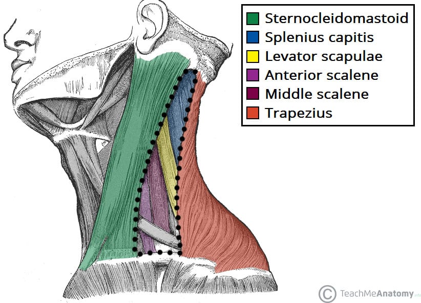
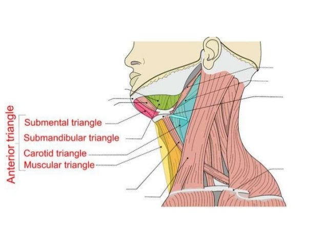
Anterior Triangle Of The Neck Subdivisions Teachmeanatomy

Jaypeedigital Ebook Reader

Jaypeedigital Ebook Reader

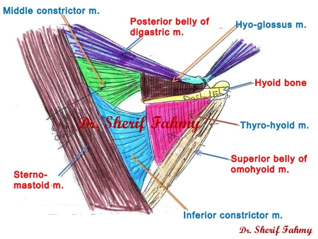
Posterior belly of digastric m.
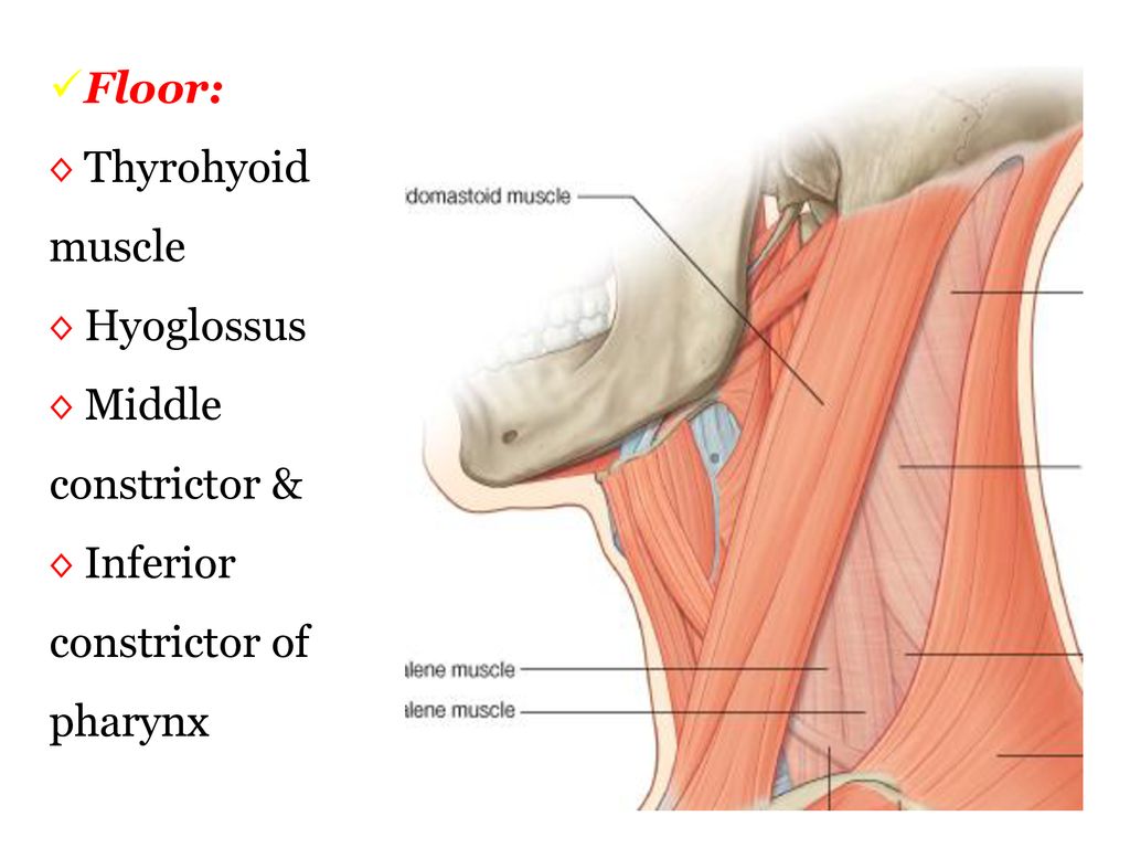
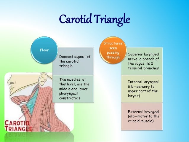
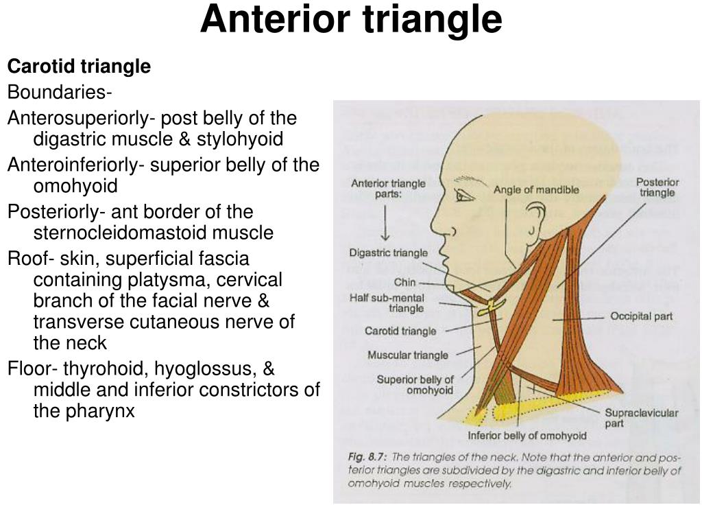
Floor of carotid triangle. Content edit edit source. However it is the posterior margin of the superior omohyoid muscle that limits the triangle anteriorly and the anterior margin of the sternocleidomastoid posteriorly. Its floor is formed by parts of the thyrohyoid membrane hyoglossus and the. Medially the floor of the triangle is formed by parts of the thyrohyoid muscle the hyoglossus muscle and the middle and inferior pharyngeal constrictor muscles.
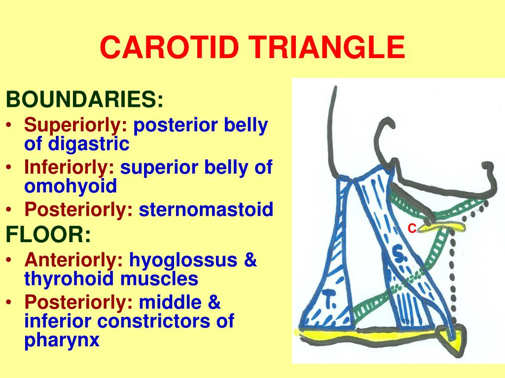
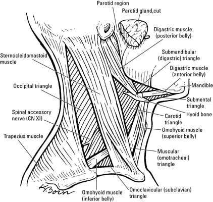
Similar to the muscular triangle the carotid triangle has the omohyoid and sternocleidomastoid muscles as parts of its borders. Floor of carotid triangle dr. Floor of digastric triangle is formed by mylohyoid anteriorly hyoglossus posteriorly infrahyoid ribbon muscles are the chief contents of muscular triangle. It is so called because it contains all the 3 carotid arteries viz.
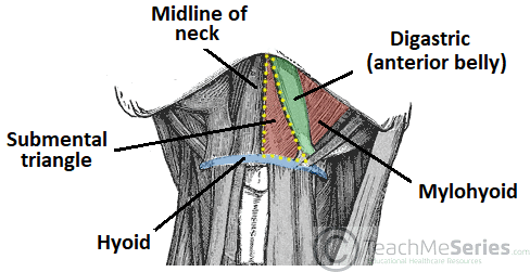
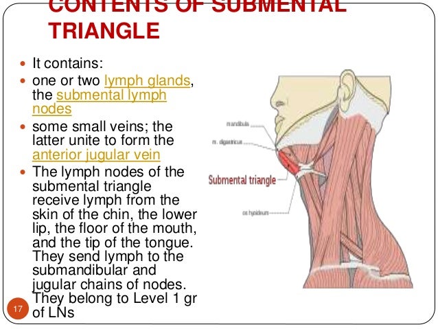
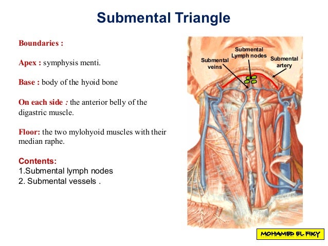
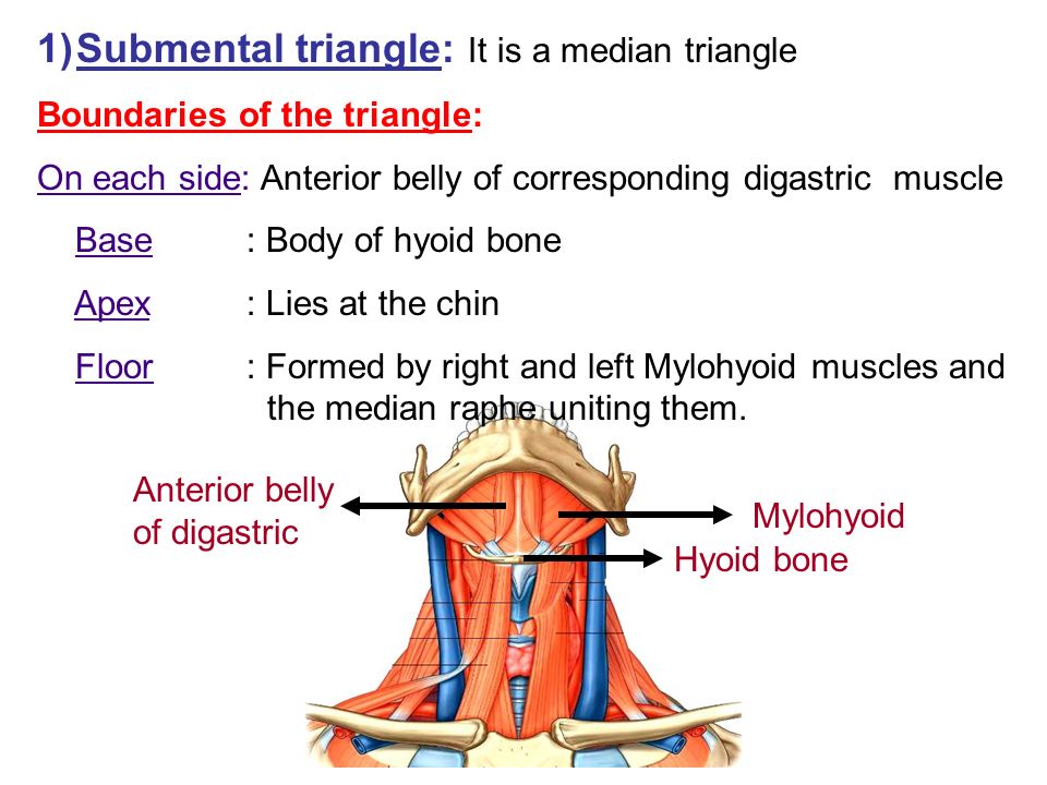
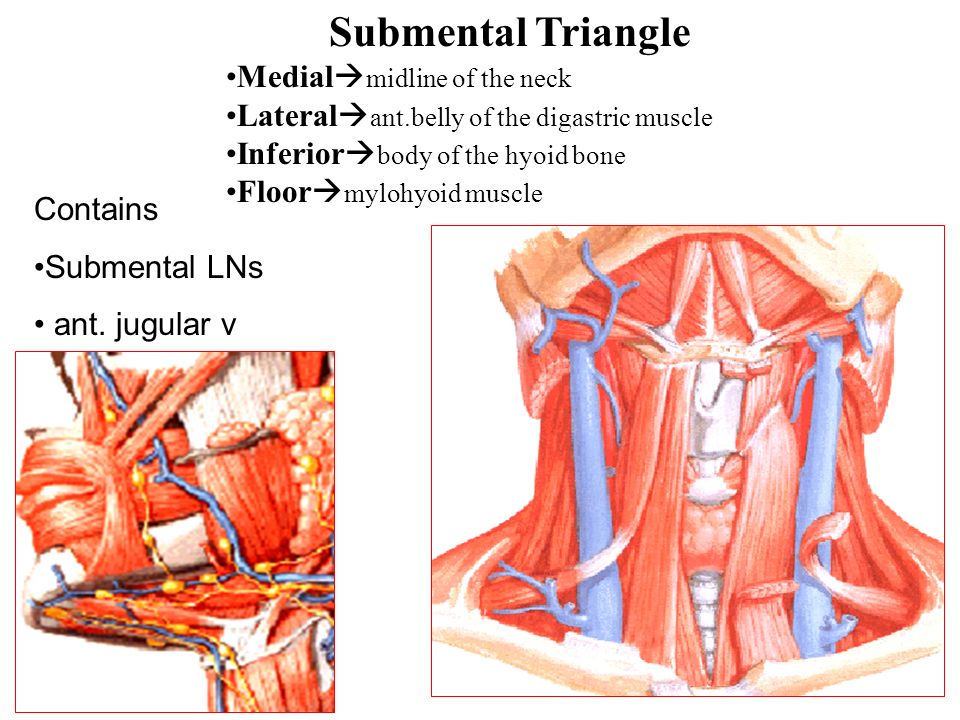
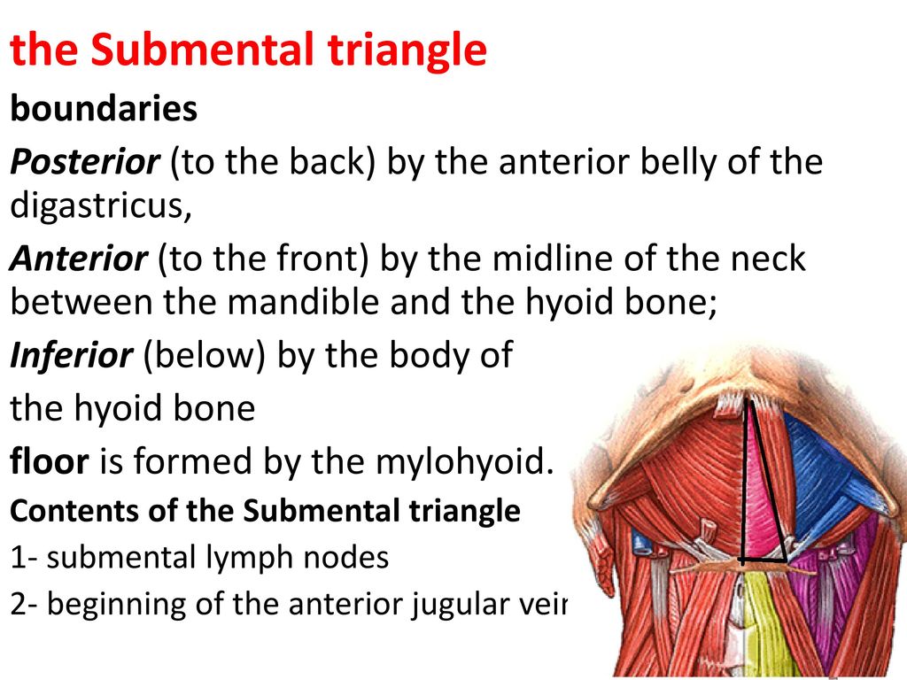
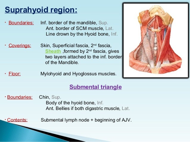
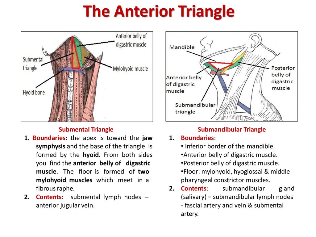
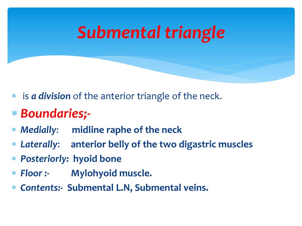
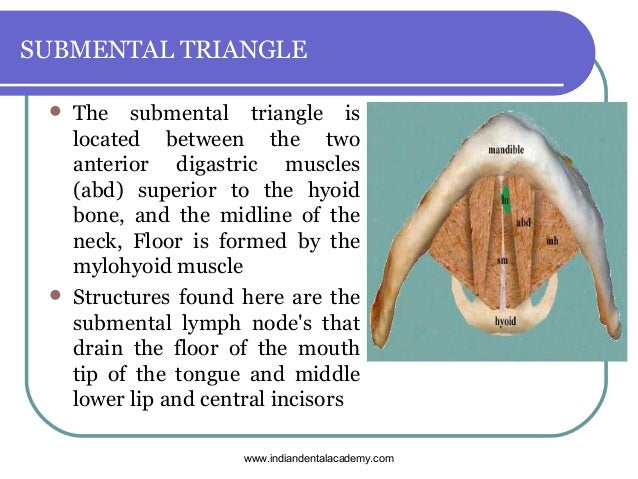
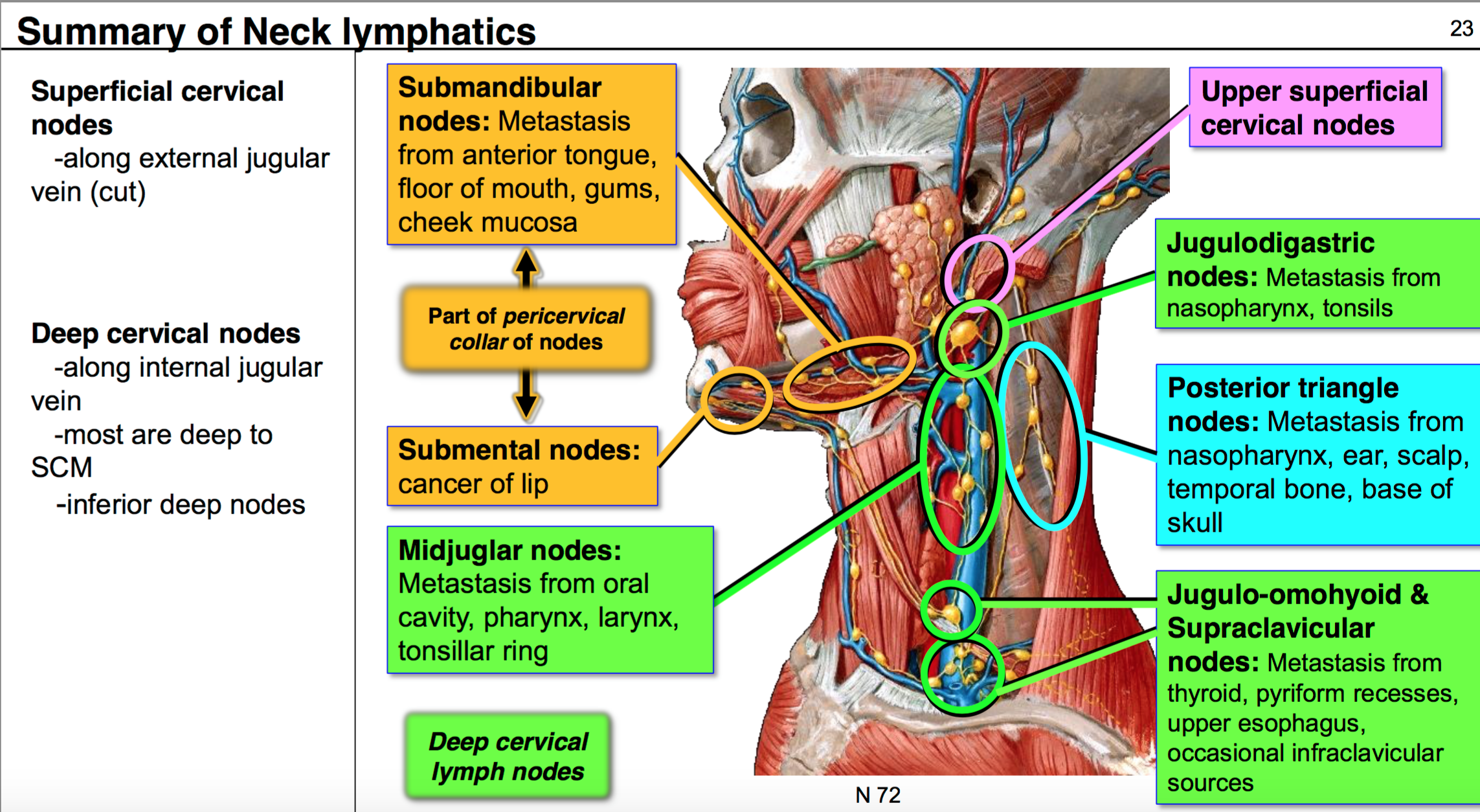
Hyoid bone thyro hyoid m. Inferiorly hyoid bone. Hyoglossus and thyrohyoid muscles posterior part. It contains the submental lymph nodes which filter lymph draining from the floor of the mouth and parts of the tongue.
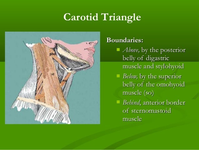
The carotid triangle or superior carotid triangle is a portion of the anterior triangle of the neck coverings and boundaries. Posteriorly by the anterior. What is the clinical significance of enlargement of the jugulo digastric and jugulo omohyoid. Carotid triangle is one of the subdivisions of anterior triangle of neck.
The carotid triangle also contains the carotid sinus a dilated portion of the common carotid and internal carotid arteries. Constrictores pharyngis medius and inferior. It is bound by the sternocleidomastoid muscle by the superior belly of the omohyoid muscle and by the posterior belly of the digastric muscle with the stylohyoideus. Common carotid internal carotid and external carotid its boundaries are.
The hyoid bone can be seen in the most anterior angle of the carotid triangle with two of the three sides either originating or inserting upon it. This triangle is situated between the ophthalmic and maxillary divisions of the trigeminal nerve and the bone of the middle fossa between the foramen rotundum and superior orbital fissure figs. Middle constrictor and inferior constrictor muscles. Superior belly of omohyoid m.
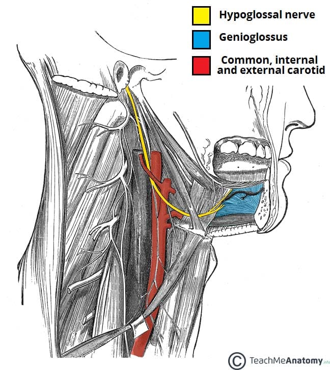
Hypoglossal nerve is a content of both digastric carotid triangles. Posterior belly of digastric and stylohyoid. Structure superficial to mylohyoid in anterior digastric triangle is mylohyoid artery nerve. Describe the locations of the superficial and deep cervical lymph nodes.
Name the structures forming the boundaries of carotid triangle. Superior belly of omohyoid. This space is used to expose the superior orbital vein and the sixth cranial nerve and to access carotid cavernous fistulae.
Carotid Triangle Radiology Reference Article Radiopaedia Org

Carotid Triangle Boundaries Contents Anatomy Tutorial Youtube
Triangles Of Neck By Dr Juveria Majeed Ms Ent

Carotid Triangle Animated Gross Anatomy Head And Neck Medical Animation Youtube
Anterior Triangle Of The Neck Part 1

Case Based Learning Triangles Of Neck Region

Anatomy Pptx د سيف Muhadharaty
Anterior Triangle Of The Neck

Contents Of Submandibular Triangle

Pdf Triangles Of The Neck A Review With Clinical Surgical Applications
Triangles Of The Neck

Triangles Of The Neck

Cb 724 Triangles Of The Head And Neck Flashcards Quizlet
B Carotid Triangle Superiorly Posterior Belly Of Digastric Ppt Download
Suboccipital Triangle Ppt Video Online Download
Stylohyoid Origin Insertion Innervation And Action Kenhub
Surgical Anatomy Of Triangles Of Neck
Triangles Of The Neck
1
The Hypoglossal Nerve Cn Xii Course Motor Teachmeanatomy
Anterior Triangle Dr Lubna Nazli Associate Professor Anatomy Ppt Video Online Download

Triangles Of Neck Flashcards Quizlet

Subclavian Triangle Wikipedia
Ppt Anterior Triangle Of The Neck Ii Powerpoint Presentation Free Download Id 5351557
Anatomy Of Neck Dr Muslim Kandel Ppt Download

Triangles Of The Neck
Unit Iv Problem Iv Anatomy Ppt Download

12 Neck Anatomy Trebloc Flashcards Quizlet

Posterior Triangle Of Neck Boundary Floor Youtube

Block 3 Anatomy Anterior Triangle Id S And Secondaries Flashcards Memorang

Digastric Triangle Boundaries And Contents Animated Gross Anatomy Head And Neck Youtube

Medicowesome Triangles Of The Neck Diagram And Mnemonic Mnemonics Medical Anatomy Anatomy And Physiology
Submandibular Triangle Anatomy And Clinical Notes Kenhub
The Posterior Triangle Of The Neck Dummies

Cervical Adenopathy And Neck Masses Radiology Key
Anterior Neck Triangles Ppt Download
Triangles Of Neck Oral Surgery Courses

Instant Anatomy Head And Neck Nerves Cranial Xi Spinal Accessory In Posterior Triangle Neck Muscle Anatomy Head And Neck Human Anatomy And Physiology

Posterior Triangle Of The Neck Ppt Video Online Download
Ppt Anterior Triangle Powerpoint Presentation Free Download Id 5436377
Anatomy G54 Neck Ii Anatomy Unit 7 Flashcards Memorang