Floor Muscles Of Digastric Triangle
Digastric Or Submandibular Triangle Earth S Lab

Anterior Triangle Of The Neck L4 Flashcards Quizlet

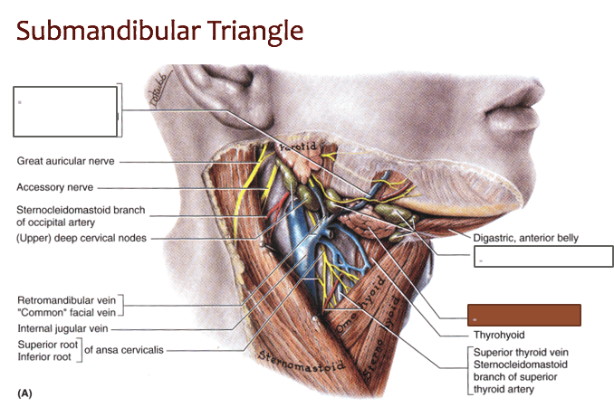
Contents Of Submandibular Triangle

Anatomy Of The Neck By Dr Rasha Sabry Ppt Video Online Download

Digastric Triangle Boundaries And Contents Animated Gross Anatomy Head And Neck Youtube

The Submandibular Gland Structure Vasculature Innervation Teachmeanatomy
The digastric muscle is composed of two bellies anterior and posterior connected by an intermediate round tendon.
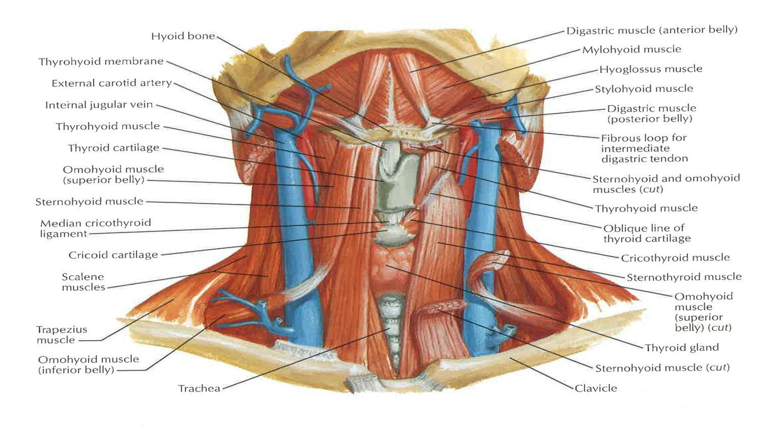
Floor muscles of digastric triangle. The carotid triangle of the neck has the following boundaries. 2 bellies of digastric anterior. Suprahyoid muscles digastric ant and post belly mylohyoid geniohyoid and stylohyoid. The anterior triangle is subdivided by the hyoid bone suprahyoid and infrahyoid muscles into four triangles.
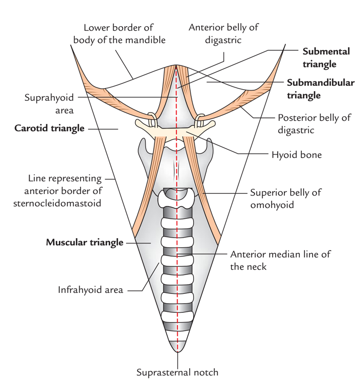
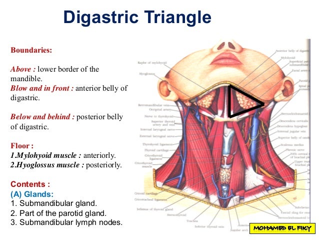
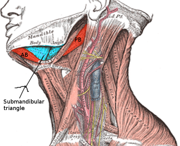
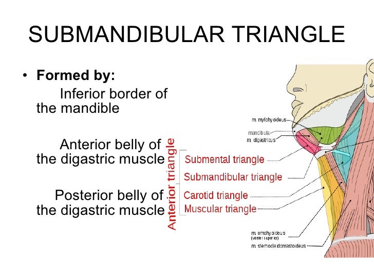
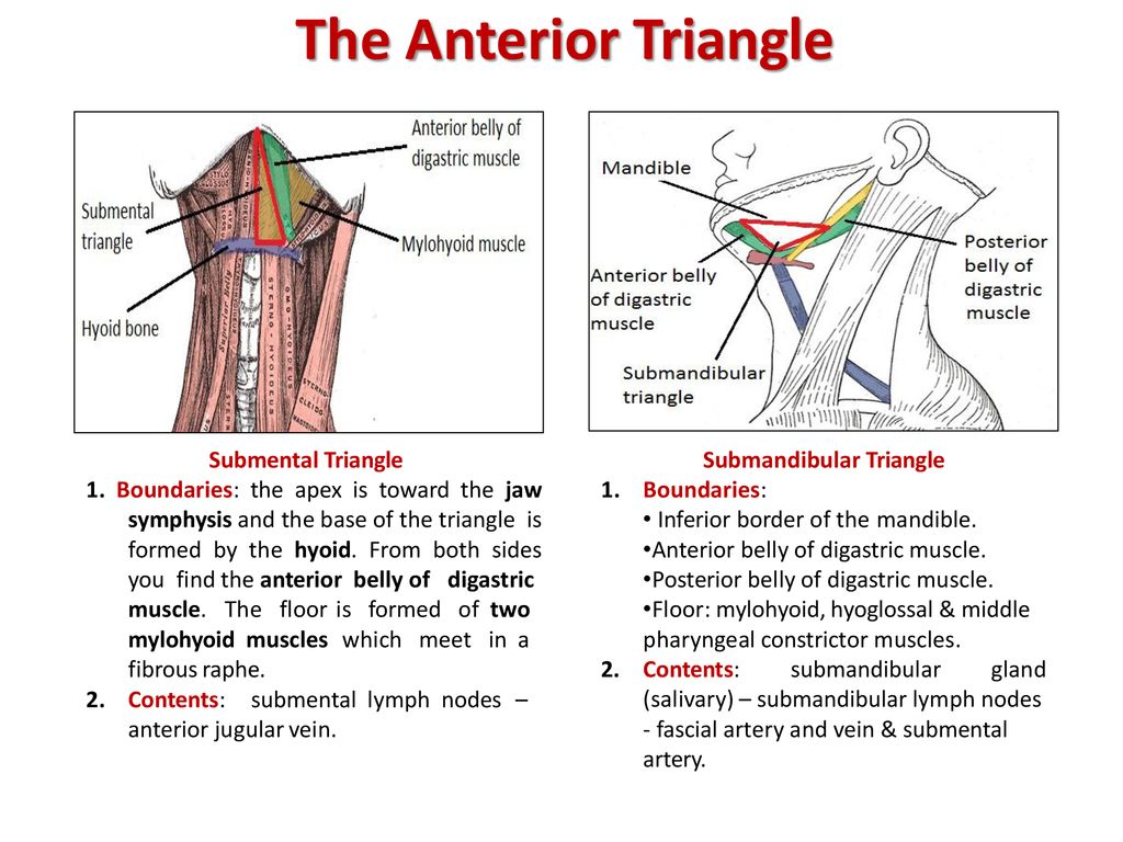
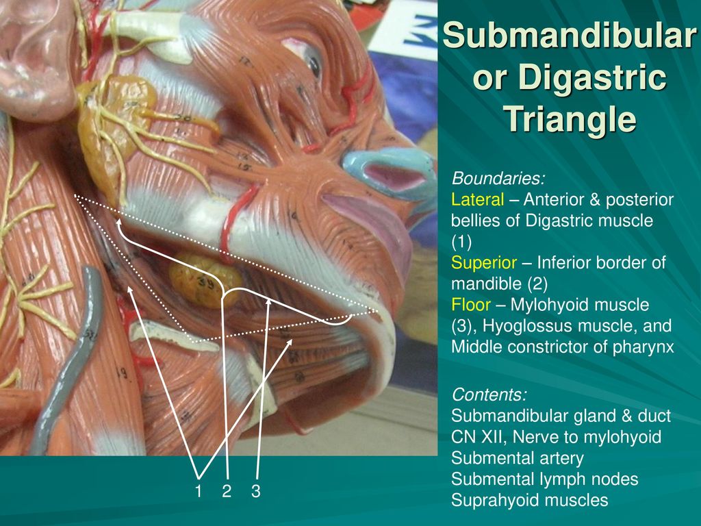
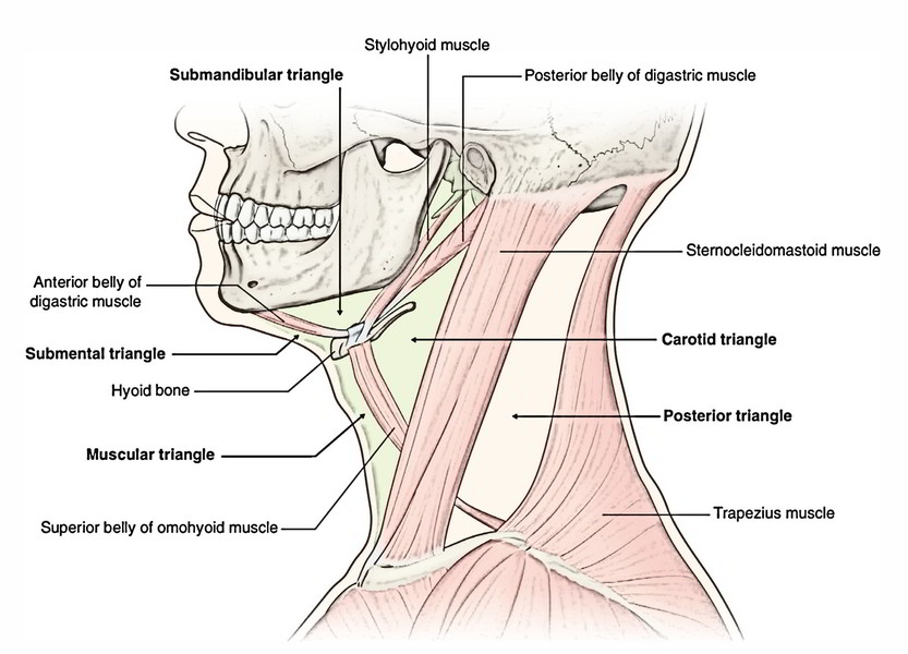
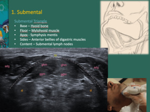
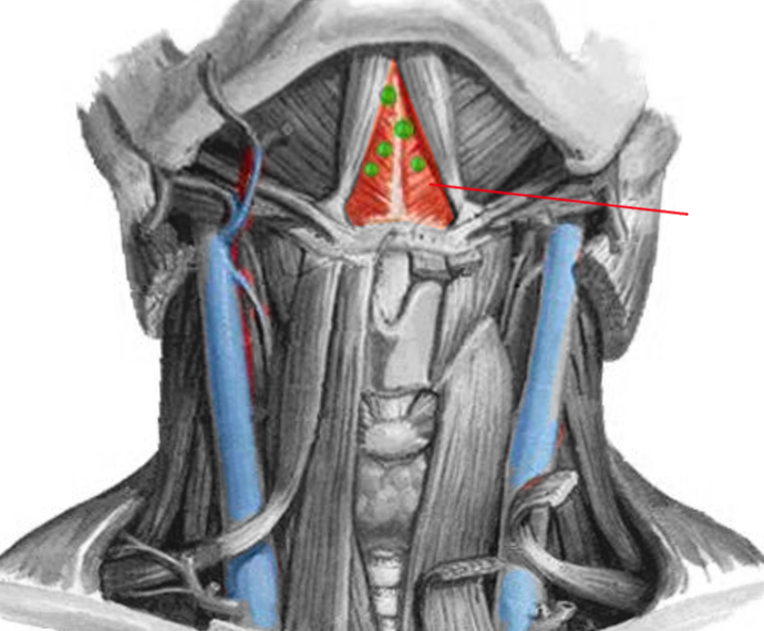
Investing fascia covers the roof of the triangle while visceral fascia covers the floor. Above by the lower border of the body of the mandible and a line drawn from its angle to the mastoid process. The digastric triangle is one of the paired triangles in the anterior triangle of the neck. Digastric triangle also known as submandibular triangle is named due to it is position in the middle of the two bellies of the digastric muscle and inferior towards the base of the mandible.
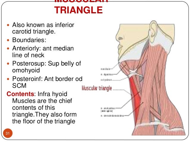
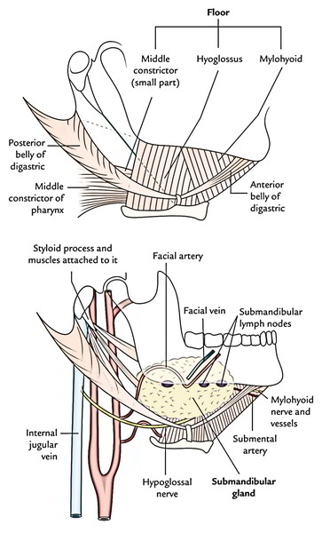
Below by the posterior belly of the digastricus. A major landmark of the submandibular triangle is the submandibular gland innervated by the facial nerve. The posterior belly of digastric muscle forms the superior border of the carotid triangle. In front by the anterior belly of the digastricus.
Superior posterior belly of the digastric muscle. What are the contents of the submandibular triangle. Muscles nerves blood vessels glands. It lies below the body of the mandible and extends in a curved form from the mastoid.
It is covered by the integument superficial fascia platysma and deep fascia ramifying in which are branches of the facial nerve and. Mastoid process of temporal bone. Infrahyoid muscles omohyoid sternohyoid sternothyroid and thyrohyoid. The two bellies of the muscle have different embryonic origins and hence are supplied by different cranial nerves.
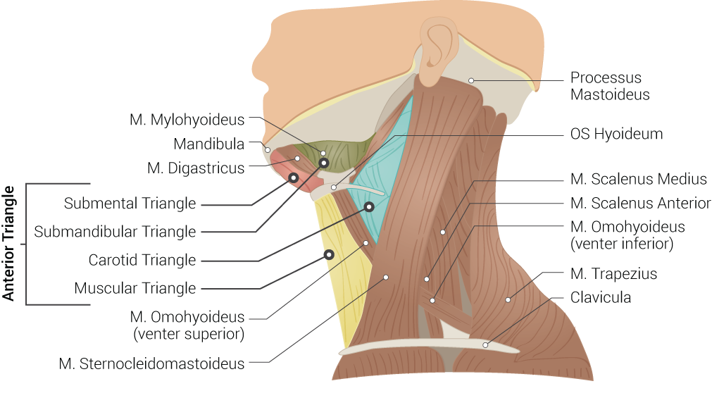
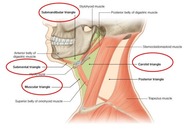
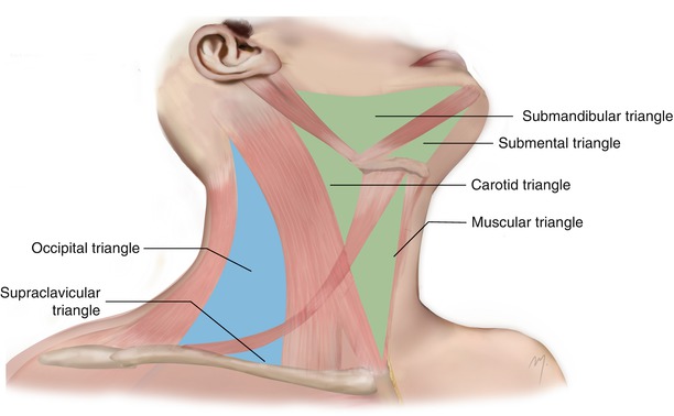
The triangles of the neck are surgically focused first described from early dissection based anatomical studies which predated cross sectional anatomical description based on imaging see deep spaces of the neck. This paired triangle contains some very important structures such as the common carotid artery internal carotid. The carotid triangle the submental triangle and the submandibular triangle. Submandibular triangle is bordered by the mandible and bellies of the digastric muscle.
On floor of mouth between mandible and genioglossus. However other muscles that have two separate muscle bellies include the ligament of treitz omohyoid occipitofrontalis. Digastric fossa on the deep surface of symphysis menti of the mandible. The digastric muscle divides the anterior triangle of the neck into three smaller divisions.
This salivary gland can be described as having two lobes which are divided by the posterior border of the mylohyoid muscle. The digastric muscle also digastricus named digastric as it has two bellies is a small muscle located under the jaw the term digastric muscle refers to this specific muscle.

Quiz6 Flashcards Quizlet

Jaypeedigital Ebook Reader
Anatomy Head And Neck Neck Triangle Article Statpearls
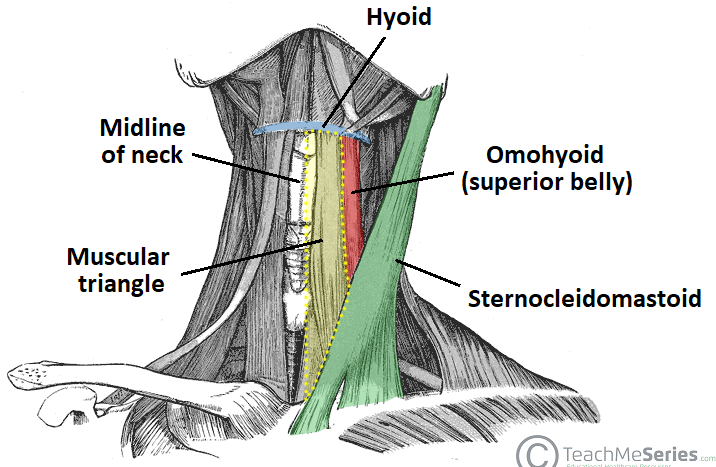
Anterior Triangle Of The Neck Subdivisions Teachmeanatomy
Anterior Triangle Of The Neck Part 1
Anatomy Head And Neck Submandibular Triangle Article Statpearls
Submandibular Triangle Anatomy And Clinical Notes Kenhub
Triangles Of Neck By Dr Juveria Majeed Ms Ent
Easy Notes On Anterior Triangle Of The Neck Learn In Just 3 Minutes Earth S Lab
Triangles Of The Neck Ppt Year 1

Neck Triangles Flashcards Quizlet

Triangles Of The Neck Anatomy Borders And Contents Kenhub

Neck Anatomy E Lab
Unit Iv Problem Iv Anatomy Ppt Download
Anterior Triangle Of Neck
Anterior And Posterior Triangles Of Neck Ppt Download

Case Based Learning Triangles Of Neck Region
001b Superficial And Deep Structures Of The Neck Anatomy Ii Flashcards Memorang
Https Encrypted Tbn0 Gstatic Com Images Q Tbn 3aand9gct Pskepy Zarbqvusgf1ltdg526fna1yyiab Fuuhyxss Lkdy Usqp Cau
Easy 3 Mins Notes On Suprahyoid And Infrahyoid Muscles Of The Neck Earth S Lab
Https Www Medicinebau Com Uploads 7 9 0 4 79048958 Anatomy Ns Note 23 Pdf
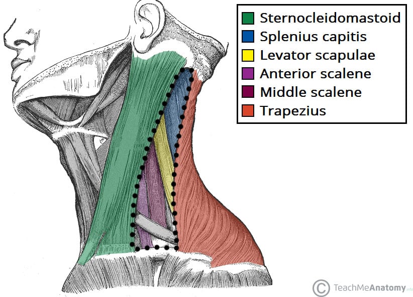
Posterior Triangle Of The Neck Subdivisions Teachmeanatomy
Neck 2 Anterior Triangle Head And Neck Anatomy Flashcards Memorang
Neck Plastic Surgery Key
Anterior Triangle Dr Lubna Nazli Associate Professor Anatomy Ppt Video Online Download

Anatomy 2 Lecture 15 And 16 Triangles Of The Neck Flashcards Quizlet

12 Neck Anatomy Trebloc Flashcards Quizlet
The Posterior Triangle Of The Neck Dummies
Triangles Of The Neck

Submandibular Triangle Download Scientific Diagram

Lab Guide 3 Anatomy For Anterior Triangle Of Neck Thyroid Gland
Triangles Of The Neck
Epos
Document 12730841

3 Anterior And Posterior Triangles Of The Neck Flashcards Quizlet
Anatomy Head And Neck Submental Triangle Article Statpearls

Triangles Of The Neck Flashcards Quizlet

Triangles Of The Neck Visual Mnemonic On Meducation

Carotid Triangle Animated Gross Anatomy Head And Neck Medical Animation Youtube

Triangle Of The Neck Davis Flashcards Quizlet

The Anterior Triangle Of The Neck Anatomy Lecture Notes

The Digastric Triangle

